


")


 – कैलकुलेटर और प्रीमियम चार्ट")

")


")
An uncommon parasitic eye infection caused by a particular kind of amoeba is known as acanthamoeba keratitis (AK). The cornea, your eye’s transparent, dome-shaped front layer, is impacted. It can harm your eyes and lead to blindness if left untreated.
There are other names for this illness, such as “amoebic keratitis.” Although it can damage both eyes, AK often affects one at a time. The epithelium, the outermost layer of your cornea, is first affected. Deeper infection is the result of worsening conditions.
Knowing about Acanthamoeba
A single-celled organism is called an amoeba. Though a little more complicated, it is comparable to bacteria. Since it can complete its life cycle without having to infect people or other animals, it is not a real parasite. However, when amoebas infect people or animals, they behave like parasites.
There are at least 20 species of acanthamoeba known to science worldwide. They easily thrive in freshwater, saltwater, soil, and a variety of other environments, practically anywhere that people do. Eight—possibly nine—acanthamoeba species are known to cause AK, according to researchers.
Two kinds are capable of acanthamoeba during their life cycles. The first is their mobile, dynamic form. Their cyst form is the other. This kind has an exterior layer that has been hardened. When in their cystic state, acanthamoeba are resistant to numerous hazards that would normally cause their death.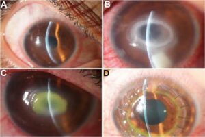
This comprises:
High temperatures: Acanthamoeba can withstand temperatures in the range of -4 to 132 degrees Fahrenheit (-20 to 56 degrees Celsius) when it is in its cyst state.
Insufficient water and nutrition: Cyst-form amebae have a 20-year (or possibly longer) shelf life at normal temperature.
Both poisons and chemicals: When in cystic form, Acanthamoeba is resistant to many chemicals’ harmful effects, including those of drugs used to treat parasite infections.
Sunlight: While many microorganisms can be killed by sunlight’s ultraviolet (UV) radiation, acanthamoeba in cyst form can survive.
It’s crucial to understand that acanthamoeba can enter your body as a cyst or an active form. You should thus take the necessary safety measures to stop them from infecting you.
Signs and Origins
What signs of keratitis caused by Acanthamoeba exist?
When these bacteria invade the corneas of your eyes in their active form, AK symptoms develop. There’s a chance that the symptoms will fluctuate, getting better and getting worse.
Among the symptoms are:
- Eye ache, occasionally quite severe.
- Feeling as though something is trapped in your eye (a foreign body sensation), but you are unable to see anything lodged there and cleaning your eyes does not alleviate the problem.
- Epiphora, or watery eyes.
- Photophobia, or sensitivity to light.
- Inflammation or redness in the eyes.
- Corneas with a ring-shaped region on their surface, foggy appearance, or dirtiness.
- Visual impairment or clouding (typically occurs with severe or advanced instances).
Acanthamoeba keratitis: what causes it?
The majority of AK cases are caused by two species of acanthamoeba (experts shorten the name of each species to “A.”). A. polyphaga and A. castellani are the two species.
Although you can contract AK from other people, it is not contagious. Instead, it is infectious.
The following are the most typical ways that acanthamoeba infects eyes:
- Lenses for contacts.
- Water that has been tainted.
- Injury to the eyes.
What side effects might acanthamoeba keratitis cause?
The following are the principal issues that could arise with AK:
- Loss of vision: Your corneas may be harmed by AK, which may result in blindness in the affected eye or eyes.
- Return: When in the form of a cyst, amethoeba can survive for a long time in your corneas and then transform back into live organisms.
- Disturbance of regular activities and routines: Usually, it’s a painful and bothersome condition. Work, hobbies, spending time with loved ones, and other activities may all be hampered by it.
Diagnoses and Examinations
How is keratitis due to canthamoeba diagnosed?
Early diagnosis of AK is challenging, with initial misdiagnosis occurring in approximately 75–90% of patients. This is due to the subsequent reasons:
- The symptoms are similar to those of more typical bacterial or viral eye diseases.
- Because AK is uncommon, eye doctors and other medical professionals typically don’t initially detect it.
- The tests need invasive corneal biopsy or scraping, as well as equipment that isn’t accessible outside of large medical facilities.
A slit-lamp examination is part of the eye exam that your eye care professional will perform. This enables them to search your eyes for indicators or clues. Inquiries on your symptoms, recent activities, and any contributing variables will also be made.
Treating any eye infection as though it were bacterial or viral first is conventional procedure. If treatment fails, it’s likely AK. In the event that it doesn’t, your eye doctor may probably advise beginning AK medication.
Tests on corneal tissue
Samples of ocular tissue may need to be taken in order to test for AK. That may entail:
- Scraping of the cornea: An eye specialist will extract a sample of the outermost layers of your cornea to analyze in order to conduct this test.
- Biopsies of the cornea: Compared to scraping, a bigger tissue sample must be taken for this. The primary benefit is that, in comparison to a scraping test, it can identify deeper infections.
These two tests have the potential to cause pain and are more intrusive. However, your provider will assist with that by using numbing drops or medicines. Additionally, corneas heal swiftly, minimizing the duration of pain or suffering following a biopsy or scrape.


")


 – सांस्कृतिक धरोहरों को नया जीवन देने की पहल")


. The cornea, your eye's)