


")


 – कैलकुलेटर और प्रीमियम चार्ट")

")


A lung disease called bronchiectasis (pronounced “bronk-ee-EK-tuh-sis”) causes damage to and enlargement of your airways, which are tubes that go to your lungs. Injured airways are unable to effectively remove mucus. After thereafter, bacteria develops in the mucus, exacerbating the inflammation and harming your lungs. You cough a lot as a result, because your body is trying to get rid of the infectious mucus.
What distinguishes bronchitis from bronchiectasis?
Coughing and lung mucus are common signs of both bronchiectasis and bronchitis. However, bronchitis is a transient infection that doesn’t leave you permanently damaged, whereas bronchiectasis causes your airways to permanently expand.
Which kinds of bronchiectasis exist?
Medical professionals classify bronchiectasis as either cylindrical (or tubular) damage to the airways, varicose damage, or cystic damage. The most prevalent and mildest type of bronchiectasis is cylindrical bronchiectasis. The most severe type is called cystic bronchiectasis.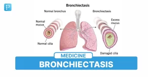
Additionally, providers classify bronchiectasis as diffuse, affecting multiple locations across the lungs, or localized, affecting just one place. The condition known as traction bronchiectasis arises when lung scarring causes your airways to become malformed.
Just how widespread is bronchiectasis?
Between 350,000 and 500,000 Americans suffer from bronchiectasis. Out of 150 individuals, one is 75 years of age or older. The true figure may be higher because bronchiectasis can occur in the absence of symptoms.
Signs and Origins
What signs of bronchiectasis are present?
Bronchiectasis symptoms include:
- Cough that is very mucusy and pussy.
- Colds that come back frequently.
- Mucus with an unpleasant scent.
- Breathing difficulty (dyspnea).
- Gasping for air.
- Spitting blood (hemoptysis) out.
- Swollen fingers and curled nails, often known as nail clubbing.
Periods of time during which your symptoms are less severe may alternate with flare-ups, or exacerbations, during which your symptoms worsen. Symptoms of an exacerbation include:
- Extreme weariness or fatigue.
- Chills and fever.
- Increased difficulty breathing.
- Sweats during night.
Why does bronchiectasis occur?
There are two stages of airway injury that lead to bronchiectasis. An infection, inflammatory disease, or other lung-related ailment causes the initial harm (or “insult”) at the first stage. Up to 40% of bronchiectasis patients have a primary etiology that is unknown to medical professionals.
Your risk of developing inflammation and recurrent infections, which worsen lung damage, increases after the initial assault. This is the “vicious cycle,” or second phase.
Diagnoses and Examinations
How is a diagnosis of bronchiectasis made?
A medical professional will examine you and inquire about your medical history in order to diagnose bronchiectasis. They will assess the function of your lungs by listening to them. To examine the anatomy of your lungs, they may prescribe imaging tests if they believe you have bronchiectasis or another lung ailment.
Which examinations are planned to identify bronchiectasis?
A medical professional may do a variety of tests to identify bronchiectasis or rule out other illnesses, such as:
Chest X-ray or CT scan: To determine whether your airways are damaged, a medical professional uses a machine to take photographs of your lungs.
Sputum cultures and blood testing: A healthcare professional obtains samples of your mucus (sputum) or blood to determine whether you have an infection.
Tests of lung function: Lung function tests are used by a clinician to assess the health of your lungs. A equipment that monitors lung function will require you to breathe into it.
Genetic examination: To check for illnesses, a healthcare professional could draw blood or other bodily fluid samples.
Test for sweat chloride: A clinician will conduct a sweat test if they suspect you may have cystic fibrosis. Your arm or leg will be made to perspire, and a sample will be taken and tested for cystic fibrosis indicators.
Bronchoscopy: In rare circumstances, a medical professional might do a bronchoscopy to have a closer look at your airways. A bronchoscope is a long, flexible tube with a light and camera at the end that is used to detect and remove anything that are obstructing your airways and to collect lung samples for testing, such as pus or mucus.


 – सांस्कृतिक धरोहरों को नया जीवन देने की पहल")


 causes damage to and enlargement of your airways, which are tubes that)