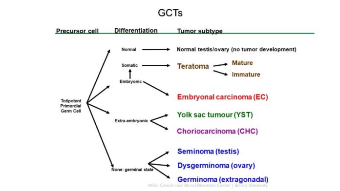
Growths of cells originating from reproductive cells are known as germ cell tumours. It’s possible that the tumors are malignant or not. The majority of germ cell tumors arise in the ovaries or testicles.
It’s unclear why some germ cell tumors can develop in other parts of the body, like the chest, abdomen, and brain. Extragonadal germ cell tumors, or germ cell tumors outside the ovaries and testicles, are extremely uncommon.
Germ cell tumors can be treated with radiation therapy using high-powered energy beams, chemotherapy using drugs that kill cancer cells, and surgery to remove the tumor.
Which kind of tumors are germ cell tumours?
Malignant (cancerous) or benign (noncancerous) germ cell tumors are both possible. Tumors of either kind have the potential to grow larger, but only cancerous germ cell tumors have the ability to spread to other body parts. Metastasized cancer is more difficult to treat and can cause damage to your organs.
Tumors containing tooth, hair, muscle, or bone are called teratomas. They could be developed or immature. The most prevalent kind of ovarian germ cell tumors are mature teratomas, also known as dermoid cysts. Usually, they are harmless. The majority of immature teratomas are malignant and grow quickly.
Cells found in yolk sac tumors, also known as endodermal sinus tumors, resemble those found in developing embryos. These tumors are malignant, and they quickly invade other organs and lymph nodes. The most frequent malignant germ cell tumor found in children is a yolk sac tumor.
Cancerous tumors called germinomas can develop in the testicles or ovaries. Nonetheless, the brain and spinal cord (central nervous system) are where they are most prevalent. If they are in your ovaries, they are known as dysgerminoma; if they are in your testicles, they are known as seminoma.
A rare kind of cancerous germ cell tumor is called embryonal cell carcinoma. It can occur in a pure form, but in a mixed germ cell tumor, it usually coexists with other tumor types.
There are parts of polyembryomas that look like embryos. These are uncommon, rapidly spreading cancerous tumors that frequently coexist with other kinds of germ cell tumors.
Cells that form the placenta during pregnancy make up choriocarcinomas. An organ called the placenta enables a gestational parent to transfer nutrients to a developing foetus. A rare and malignant germ cell tumor, choriocarcinoma typically develops in the uterus but can also arise in the testicles or ovaries. Both the parent and the foetus may contract it.
Two or more types of malignant germ cell tumors can coexist in mixed germ cell tumors. Germ cell tumors are often heterogeneous.
Causes
In a human embryo in development, normal germ cells form. These cells eventually develop into egg or sperm cells when they reach the embryo’s ovaries or testicles.
On the other hand, cells that do not mature into fully developed eggs or sperm make up germ cell tumors. Instead, the germ cells divide erratically and grow into a tumor in your testicles or ovaries. Extragonadal tumors occur when germ cells invade unusual body locations, such as the chest, brain, belly, low back, and tailbone, and grow into tumors.
It’s unclear to researchers why germ cells develop in this manner.
Symptoms
The size and location of the tumor in your body are important factors that affect how a germ cell tumor feels.
Tumours of ovarian germ cells
Not all ovarian germ cell tumors present with symptoms. Mature teratomas, for example, might not show symptoms until they’ve gotten big enough to put pressure on your abdomen. They are frequently found during an ultrasound to identify the source of pelvic pain.
Among the symptoms could be:
- pain or discomfort in the pelvis.
- ovarian mass that hurts.
- enlarged abdomen, or belly.
- stomach ache (akin to appendicitis).
- irregular bleeding in the vagina.
- nausea.
Tumours of testicular germ cells
Testicular cancer and testicular germ cell tumors present with similar symptoms.
Among the symptoms are:
- a firm, solid lump that enlarges in the testicle (with or without pain).
- feeling achy or heavy in your crotch.
- either groyne or abdominal pain.
- strangely shaped testicle.
- back discomfort.
Tumours of extragonadal germ cells
A teratoma may be indicated by a mass in the middle of your child’s chest or on their tailbone. Depending on where the extragonadal germ cell tumor is located, there may be additional symptoms.
- breathing difficulties (lungs).
- Leg weakness (low back).
- Urinating and defecating (pelvis).
- Edoema and either severe or mild abdominal pain in your child.
Diagnosis
Along with a physical examination, your healthcare provider will inquire about your symptoms. To identify a germ cell tumor, they could carry out any of the following examinations or procedures:
CT scan: A CT scan generates a computerised, three-dimensional image of your soft tissues and bones by combining several interior X-ray pictures. The location of a tumor can be seen with a CT scan of your chest, abdomen, or pelvis.
MRI: An MRI produces a computerised image of your body’s soft tissue and bone using radio waves and magnets. An MRI can display the location of your tumor, just like a CT scan.
Ultrasound: An ultrasound produces images of your interior organs by using sound waves. The amount of a tumor that is a solid mass or cystic (fluid-filled) can be determined by ultrasound. When trying to diagnose a growth as either a cyst or a germ cell tumor, this information can be useful.
PET scan: A tracer used in a PET scan can reveal the location of cancer cells within your body. If cancer has spread, a PET scan can reveal it.
Bone scans: An X-ray of your bones is taken after they have absorbed a particular dye that highlights any abnormalities. If you have a tumor affecting your bones, bone scans can reveal it.
Blood tests: To measure the levels of proteins, hormones, or enzymes in your blood, your doctor may perform a blood draw. Certain types of germ cell tumours may be indicated by elevated levels of lactate dehydrogenase, AFP, and human chorionic gonadotropin (hCG).
Biopsy: During a biopsy, tissue from the tumor is removed and sent to a lab for analysis. To identify a germ cell tumor, a specialist known as a pathologist examines the sample under a microscope.


